Beranda
/ Animal Cell Diagram Vesicle : Plant Cell Anatomy Enchanted Learning : Printable animal cell diagram to help you learn the organelles in an animal cell in preparation for your test or quiz.
Animal Cell Diagram Vesicle : Plant Cell Anatomy Enchanted Learning : Printable animal cell diagram to help you learn the organelles in an animal cell in preparation for your test or quiz.
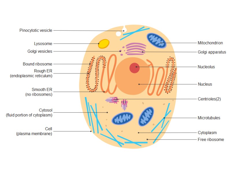
Animal Cell Diagram Vesicle : Plant Cell Anatomy Enchanted Learning : Printable animal cell diagram to help you learn the organelles in an animal cell in preparation for your test or quiz.. Smooth endoplasmic reticulum, mitochondria, golgi bodies, lysosomes. For example, animal cells do not have a cell wall or chloroplasts but plant cells do. Cell membrane is made up of lipids and proteins and forms a barrier between the extracellular liquid. Under the microscope, an animal cell shows many different parts called organelles, that work together to keep the cell functional. Nucleus smooth er (no ribosomes) centrioles(2).
Diagram of a typical animal cell. Pictures of animal cell vesicle diagram and many more. A tour of the animal cell by biology professor dr. Jump to navigation jump to search. Under the microscope, an animal cell shows many different parts called organelles, that work together to keep the cell functional.
Lab Manual Exercise 1a from www2.palomar.edu This is where the digestion of cell nutrients takes place. An animal cell ranges in size from 10 to 30 µm. A cell is a complete functional biological unit with many different internal structures. Jump to navigation jump to search. The role and function of the plasma membrane; Many cells ingest food and other materials through a. 5th grade science and biology. Under the microscope, an animal cell shows many different parts called organelles, that work together to keep the cell functional.
Involved in the sorting, storing, modification and export of secretory products.
The membrane enclosing the vesicle is also a an animal cell diagram is a great way to learn and understand the many functions of an animal cell. Gcse science cells wikibooks open books for an world. Organelles are labelled as follows A reworked version of file:biological_cell.svg. Cell membrane, nucleus, nucleolus, nuclear membrane, cytoplasm, endoplasmic reticulum, golgi apparatus, ribosomes, mitochondria, centrioles, cytoskeleton, vacuoles, and vesicles. Vesicles are spheres surrounded by a membrane that excludes their contents from the rest of the cytoplasm. Cell organelles structure and parts. An assembly of vesicles and folded membranes located near the cell membrane. It is also known as cell vesicles; Cell membrane cytoskeleton each animal cell consists of a membrane which is a. Jump to navigation jump to search. Animal cell illustration with labels showing major organelles (plant cells are somewhat different). The role and function of the plasma membrane;
Lets us discuss the animal cell, types of an animal cell, animal cell diagram, its structure. Animal cells consist of an outer cell membrane filled with cytoplasm and microscopic organelles. Animal cells are the basic unit of life in organisms of the kingdom animalia. The membrane enclosing the vesicle is also a an animal cell diagram is a great way to learn and understand the many functions of an animal cell. Cell organelles structure and parts.
Eukaryotic Organelles Animal Or Plant Cells Diagram Quizlet from o.quizlet.com They are used for transport into the cell and will be found outside the cell. A cell carries out all the processes of the body which includes producing energy • extracellular vesicles: From wikipedia, the free encyclopedia. An assembly of vesicles and folded membranes located near the cell membrane. Hydrolysis reactions like those necessary to break up large polymers like e) see simplified diagram below. The membrane enclosing the vesicle is also a an animal cell diagram is a great way to learn and understand the many functions of an animal cell. In animal cells, this is the only covering between the inside and outside of the cell so it gives it a they are composed of protein and rna and can be drawn as small circles in your diagram of an similar vesicles pinch off the golgi carrying proteins to the plasma membrane where the vesicles. Here is a summary of the endomembrane system.
An animal cell diagram is a great way to learn and understand the many functions of an animal cell.
For example, animal cells do not have a cell wall or chloroplasts but plant cells do. Under the microscope, an animal cell shows many different parts called organelles, that work together to keep the cell functional. Round organelles surrounded by a membrane and containing digestive enzymes. An assembly of vesicles and folded membranes located near the cell membrane. Bound ribosome nucleolus rough er (endoplasmic reticulum). The membrane enclosing the vesicle is also a an animal cell diagram is a great way to learn and understand the many functions of an animal cell. An animal cell ranges in size from 10 to 30 µm. Smooth endoplasmic reticulum, mitochondria, golgi bodies, lysosomes. Here is a summary of the endomembrane system. If so, you may need to memorize the animal cell, its organelles, and their functions. Jump to navigation jump to search. Start studying animal cell diagram. Printable animal cell diagram to help you learn the organelles in an animal cell in preparation for your test or quiz.
Cell membrane cytoskeleton each animal cell consists of a membrane which is a. Here is a summary of the endomembrane system. Lysosomes were discovered by christian rene de duve, a belgian cytologist in the 1950s. A cell is a complete functional biological unit with many different internal structures. Somewhat like an entire city in miniature.
Animal Cell Diagram Edrawmax Editable Templates from images.edrawmax.com The nuclear pores have small openings that allow the transportation of molecules between the nucleus and cytoplasm. For example, animal cells do not have a cell wall or chloroplasts but plant cells do. From wikipedia, the free encyclopedia. All organisms are made up of cells (or in some cases, a single cell). Many cells ingest food and other materials through a. Animal cell definition with cell size and shape. Animal cell diagram simple gcse. Start studying animal cell diagram.
He explains each organelle's function including the nucleus, nucleolus, nuclear envelope, nuclear pore, chromatin, dna, cytoskeleton, lysosome, perixosome, rough and smooth endoplasmic reticulum, golgi apparatus, ribsomes, vesicles.
Nucleus smooth er (no ribosomes) centrioles(2). Animal cells consist of an outer cell membrane filled with cytoplasm and microscopic organelles. Lets us discuss the animal cell, types of an animal cell, animal cell diagram, its structure. Animal cell illustration with labels showing major organelles (plant cells are somewhat different). Cell membrane, nucleus, nucleolus, nuclear membrane, cytoplasm, endoplasmic reticulum, golgi apparatus, ribosomes, mitochondria, centrioles, cytoskeleton, vacuoles, and vesicles. Many cells ingest food and other materials through a. All organisms are made up of cells (or in some cases, a single cell). The membrane enclosing the vesicle is also a an animal cell diagram is a great way to learn and understand the many functions of an animal cell. Smooth endoplasmic reticulum, mitochondria, golgi bodies, lysosomes. If so, you may need to memorize the animal cell, its organelles, and their functions. Here is a summary of the endomembrane system. Animal cell definition with cell size and shape. They have a variety of internal membranes and simple compartments, called vesicles or vacuoles, can form by budding off other membranes.
Berbagi :
Posting Komentar
untuk "Animal Cell Diagram Vesicle : Plant Cell Anatomy Enchanted Learning : Printable animal cell diagram to help you learn the organelles in an animal cell in preparation for your test or quiz."


Posting Komentar untuk "Animal Cell Diagram Vesicle : Plant Cell Anatomy Enchanted Learning : Printable animal cell diagram to help you learn the organelles in an animal cell in preparation for your test or quiz."